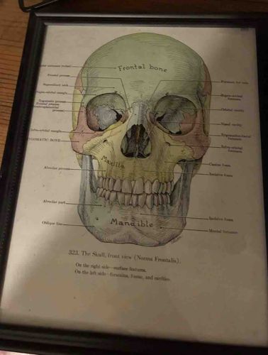
Framed Anatomical Illustration of the Human Skull (Norma Frontalis)
Vintage Medical Illustration / Art Print

Framed Anatomical Illustration of the Human Skull (Norma Frontalis)

This item is a color-coded anatomical print depicting the human skull from a frontal view, titled '323. The Skull, front view (Norma Frontalis)'. The illustration is highly detailed, featuring specific labels for various cranial structures including the Frontal bone, Maxilla, Mandible, and Zygomatic bone. The color palette utilizes muted tones of sage green, yellow-ochre, and reddish-tan to differentiate the various sections of the skull, a common technique in medical textbooks from the late 19th to early 20th centuries. The text below the primary image explains that the right side identifies surface features while the left side identifies foramina, fossae, and cavities. The print is housed in a contemporary black wood or composite frame with a beaded inner edge, indicating it has been preserved or displayed as decorative art. The paper shows a slight warm patina consistent with age, though no significant tears or foxing are visible through the glass. The typography and fine-line etching style suggest this may be an original book plate removed from a classic medical atlas such as Gray's Anatomy or a similar reference work from the vintage period. Overall, the piece displays high-quality scientific craftsmanship and remains in excellent condition with clear, legible text and vibrant, un-faded color pigments.

Estimated Value

$150-$225

Basic Information

Category

Vintage Medical Illustration / Art Print

Appraised On

January 19, 2026

Estimated Value

$150-$225

Item Description

This item is a color-coded anatomical print depicting the human skull from a frontal view, titled '323. The Skull, front view (Norma Frontalis)'. The illustration is highly detailed, featuring specific labels for various cranial structures including the Frontal bone, Maxilla, Mandible, and Zygomatic bone. The color palette utilizes muted tones of sage green, yellow-ochre, and reddish-tan to differentiate the various sections of the skull, a common technique in medical textbooks from the late 19th to early 20th centuries. The text below the primary image explains that the right side identifies surface features while the left side identifies foramina, fossae, and cavities. The print is housed in a contemporary black wood or composite frame with a beaded inner edge, indicating it has been preserved or displayed as decorative art. The paper shows a slight warm patina consistent with age, though no significant tears or foxing are visible through the glass. The typography and fine-line etching style suggest this may be an original book plate removed from a classic medical atlas such as Gray's Anatomy or a similar reference work from the vintage period. Overall, the piece displays high-quality scientific craftsmanship and remains in excellent condition with clear, legible text and vibrant, un-faded color pigments.

Get Your Items Appraised

Instant estimates of your treasures with AI-powered instant appraisals

Browse More Appraisals